Spectroscopy
IR spectroscopy
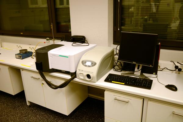
FT-IR spectrometer (Nicolet Avatar 360) was installed in 2003. The spectral range of the instrument is 7800-375 cm-1 and it is using proprietary K Br beam splitter. Omnic software is used for data collection and processing.
Raman spectroscopy
Raman spectrometer
The spectrometer (The dispersive Kaiser Raman Hololab series 5000) is equipped with 785 nm laser. The spectrometer can be connected to the additional non-microscopic probehead with 1.5 and 20m optical cables. The long, 20 m optical cable enables the in situ measurements even in industrial environment. The system is also equipped with Olympus microscope which makes possible micro-Raman analyses, Raman mapping and confocal Raman measurements. The system is controlled with Holograms software and data processing is made with GRAMS32 software.
UV Resonance Raman spectrometer
The Raman spectrometer (Renishaw 1000 UV) equipped with microscope of 15X and 40X objectives. The laser is Innova 300C FreDTM frequency doubled Ar+ Ion Laser (Coherent, Inc., California), which is operated at three different wavelengths (229, 244 and 257 nm). CCD camera is used for detecting the scattered light. The system is controlled and the data is processed with Grams32 software.
The method is mainly used for analysing residual lignin structures from pulp samples. The Raman signals of aromatic and conjugated lignin structures are enhanced by resonance effect with the UV excitation, whereas other wood components are not enhanced. Therefore, lignin characterization is possible even from fully bleached pulps. Measurements last only about 30 seconds per sample. Additionally, the measurements are accomplished directly from department hand sheets and pretreatment of samples is not needed.
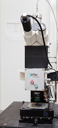
AFM-Raman spectrometer
WITec alpha 300 RA combined Raman microscope and atomic force microscopehas possibility to perform Confocal Raman mapping and AFM imaging.
Details about Raman:
- Excitation laser: Frequency doubled Nd:YAG green (λ=532.25 nm, 30 mW)
- Objectives: Olympus 100X (NA=0.95) and 20X (NA=0.4) air objectives and 10X and 60X liquid objectives
- Detector: Electron multiplying EMCCD camera (Andor Newton DU970-BV)
- Half-wave plate placed in the optical pathway to perform orientation experiments.
Details about AFM:
- All basic imaging modes are available (contact, AC, phase detection, lateral force mode...)
- Fluid cell available for measurements in liquid
- Pulsed force mode available
- The instrument is equipped with active vibration insulation system (TS-150).
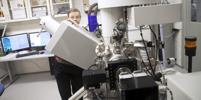
X-ray photoelectron spectroscopy (XPS)
XPS, X-ray photoelectron spectroscopy; also called as ESCA, Electron spectroscopy for chemical analysis (AXIS Ultra by Kratos Analytical) is used in chemical characterization of the topmost 10 nanometres of solid materials. Sample is bombarded with soft X-rays in ultra-high vacuum.
- Quantitative elemental composition at the surface (all but H, He are detected)
- Information about chemical compounds at the surface, based on nearest neighbours and oxidation states
- Surface distributions and modelling via spectral background evaluation within 10 nm
- Surface profiling via sputtering
- NEW: Surface chemical mapping (lateral resolution under 10 micrometers)
- NEW: CryoXPS, including analyses of wet, frozen surfaces at -150 °C.
- The new state-of-art XPS instrument with a monochromatic irradiation and effective neutraliser was installed in 2013. It is customised for soft materials. However, the instrument can be utilised in many other surfaces studies too, e.g. in contamination & failure analyses; characterisation of thin films, composites and heterogeneous surface structuresas well as in metallurgy and polymer science.
- Examples of research conducted for cellulosic materials at Aalto: determination of surface lignin and extractives from hand sheets, surface analyses on papers, textiles, composite materials, grafted celluloses, nanocelluloses, natural fibres and wood.
- Inquiries: Dr. Leena-Sisko Johansson: leena-sisko.johansson(at)aalto.fi
Atomic-force microscopy (AFM) - Force spectroscopy
AFM: The NanoScope MultiMode scanning probe microscope (SPM) consists of the MultiMode SPM, NanoScope IIIa control system and NanoScope image analysis and presentation software (version 4.43r8 for Windows NT). The system includes the Extender TM Electronics Module making phase imaging possible in addition to the other available modes (contact AFM, Tapping Mode AFM, force modulation, and lateral force microscopy). The apparatus is equipped with tip holders for measurements in air and liquid and a 125 µm as well as a 12 µm scanner. The system includes a 45X monocular microscope and an optical microscope with zoom, binocular, black & white camera and monitor, making accurate positioning on the sample easy.
UV spectroscopy
The department has routine instruments for UV spectroscopy.
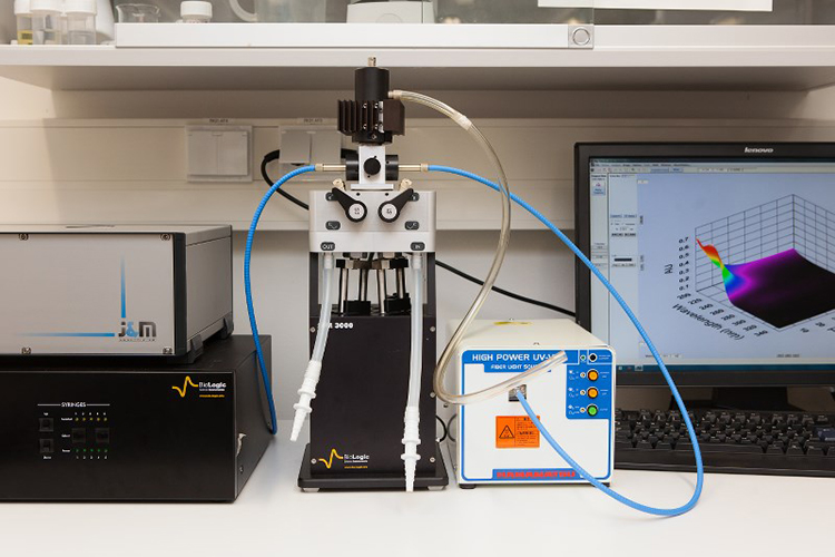
Stopped flow device (SFM-3000, Bio-Logic): stopped-flow module (SFM) consists of a mechanical subsystem and a motor power supply. In the SFM-3000, the mechanical sub-system consists of three machined syringes and one valve block with 3´3-way valves, with the possibility to include one or two mixers and one ageing loop. All SFM syringes, valves, delay lines, and cuvettes are enclosed in a water jacket to allow temperature regulation of the reactants’ containers.
Fluorescence Spectroscopy
The Cary Eclipse Spectromether uses a Xenon flash lamp for superior sensitivity, high signal-to-noise ratio and fast kinetics. Four collection modes: Fluorescence, phosphorescence, chemi/bioluminescence and time resolved phosphorescence.
Other equipment
Refractometer (Abbemat 300 Anton Paar) is for measuring the refractive index in harsh environments and for special applications.
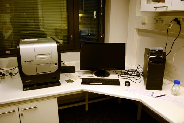
The Cytation 3 Cell Imaging Multi-Mode microplate reader combines automated digital microscopy and conventional microplate detection. Detection modes: UV-Vis absorbance, fluorescence intensity, luminiscence.
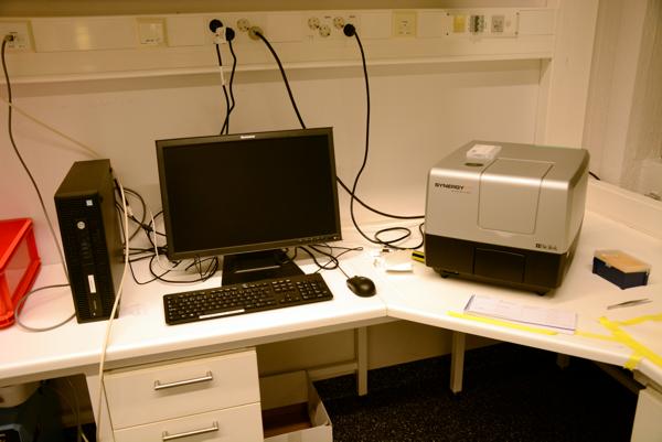
The Synergy H1 Multi-Mode Reader is a microplate reader for conventional microplate detection. Detection modes: UV-Vis absorbance, fluorescence intensity, luminiscence.